
A Bioinformatic Investigation into the Role of the CXCR4 Chemokine Receptor in Cancer Progression and Metastasis: Expression, Prognosis, and Immune Infiltration
Project Overview: The chemokine receptor CXCR4 and its cognate ligand CXCL12 (SDF-1) regulate tumor cell migration, invasion, and organ-selective metastasis. This thesis performed a multi-cancer in silico investigation of CXCR4 across Breast Invasive Carcinoma (BRCA), Lung Adenocarcinoma (LUAD), and Colorectal Adenocarcinoma (COAD/READ) using genomic, transcriptomic, and clinical data from The Cancer Genome Atlas (TCGA). The study evaluated differential mRNA expression, copy-number alterations, overall survival, CD8+ T-cell infiltration, and protein–protein interaction (PPI) networks, revealing that CXCR4 demonstrates context-dependent prognostic behavior that challenges the assumption of a universally adverse biomarker.
Research Questions & Hypotheses
The study evaluated whether the biological behavior and clinical significance of CXCR4 conform to a uniform oncogenic paradigm or whether they are governed by tissue-specific microenvironments:
- Differential Expression: Is CXCR4 mRNA expression significantly elevated in tumor tissues compared to matched normal tissues across breast, lung, and colorectal cancers?
- Genomic Drivers: Is elevated CXCR4 expression driven by somatic structural genomic alterations (such as gene amplifications or mutations) or by transcriptional and microenvironmental dysregulation?
- Prognostic Divergence: Does high CXCR4 expression consistently correlate with reduced patient overall survival across different cancer cohorts?
- Microenvironment Correlation: Is CXCR4 expression correlated with immune cell infiltration—specifically cytotoxic CD8+ T lymphocytes—in tumors where survival associations diverge?
Working Hypotheses: (H1) CXCR4 is overexpressed across solid tumors predominantly through transcriptional mechanisms rather than genomic amplification. (H2) High CXCR4 expression is associated with poor prognosis, but this relationship is modulated by the immune infiltration state of the local tumor microenvironment.
Bioinformatic Methodology
The analysis was conducted across standardized clinical cohorts from The Cancer Genome Atlas (TCGA) using five validated bioinformatic analytical platforms:
Expression Profiling
Utilized GEPIA2 to assess CXCR4 mRNA expression in tumor samples versus TCGA and GTEx matched normal tissues in BRCA, LUAD, and COAD. Statistical significance evaluated with one-way ANOVA (cutoffs: Log2FC = 1, p < 0.01).
Genomic Alterations
Queried TCGA Pan-Cancer Atlas datasets on cBioPortal for Breast, Lung, and Colorectal cohorts. Evaluated mutation frequency, deep deletions, and putative copy-number status against mRNA abundance.
Survival Analysis
Assessed Overall Survival (OS) using Kaplan-Meier Plotter in BRCA, LUAD, and READ cohorts. Log-rank test and Hazard Ratios (HR) with 95% Confidence Intervals calculated with software-determined optimal cutoff values.
Immune & PPI Networks
Analyzed correlation between CXCR4 expression and CD8+ T-cell infiltration via TIMER2.0 using Spearman's Rho across multiple deconvolution algorithms. Built protein–protein interaction networks via STRING.
Data Sources & Platforms
Key Findings & Context-Dependent Prognosis
1. Significant Overexpression in Breast & Colon; Modest in Lung
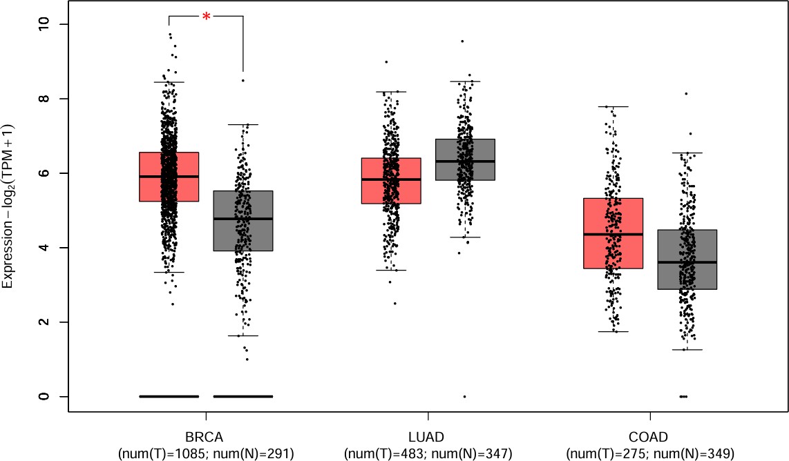
GEPIA2 analysis showed that CXCR4 mRNA expression was significantly higher in tumor tissues than in normal controls in Breast Invasive Carcinoma (BRCA) and Colon Adenocarcinoma (COAD) (p < 0.01). In contrast, Lung Adenocarcinoma (LUAD) exhibited only a modest, non-significant difference between tumor and matched normal tissues.
2. Transcriptional/Microenvironmental Drivers Over Structural Genomic Amplification
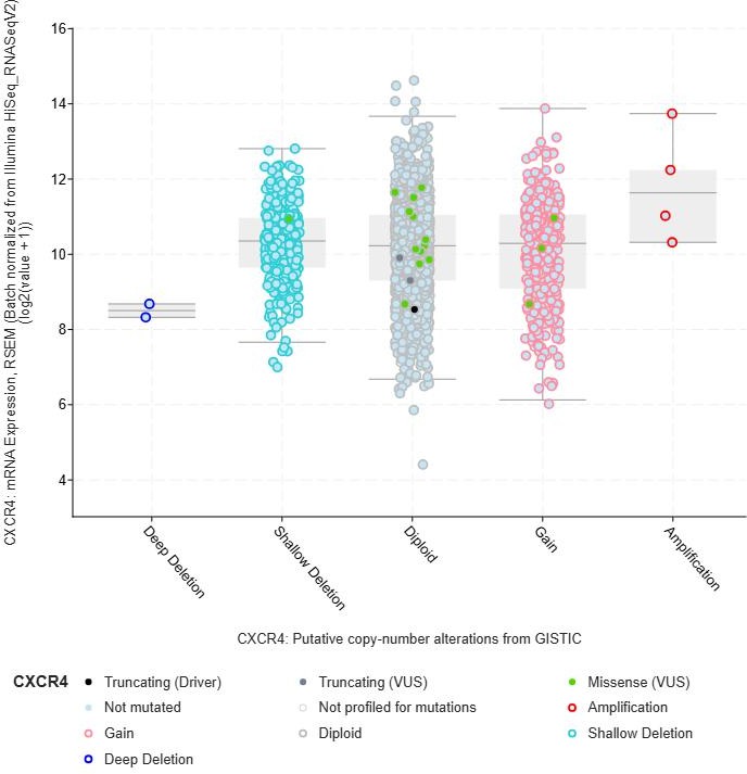
Somatic mutations and copy-number amplifications in CXCR4 were uncommon across the queried TCGA cohorts (<2%). Although amplified samples generally showed high expression, a large proportion of tumors in the "Diploid" group (normal copy number) also displayed elevated mRNA abundance. This indicates that genomic amplification does not drive CXCR4 overexpression; rather, transcriptional or microenvironmental induction (e.g., hypoxia-mediated HIF-1α signaling) represents the primary mechanism.
3. The LUAD Prognostic Paradox (p = 0.007)
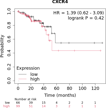
Survival outcomes differed across tissues. In BRCA and READ, CXCR4 expression showed no statistically significant association with Overall Survival (p > 0.05). In striking contrast, in Lung Adenocarcinoma (LUAD), higher CXCR4 expression was significantly associated with longer overall survival (p = 0.007).
4. Correlation with CD8+ T-Cell Infiltration
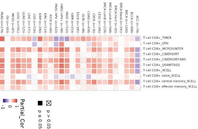
TIMER2.0 analysis revealed a consistent positive correlation (Spearman's Rho > 0) between CXCR4 expression and CD8+ cytotoxic T-cell infiltration in LUAD across multiple algorithms. Rather than proving direct causation, this association suggests that high CXCR4 expression in LUAD may serve as an indirect surrogate marker for an immune-enriched, immunologically active tumor microenvironment.
5. PPI Chemokine Network Connectivity
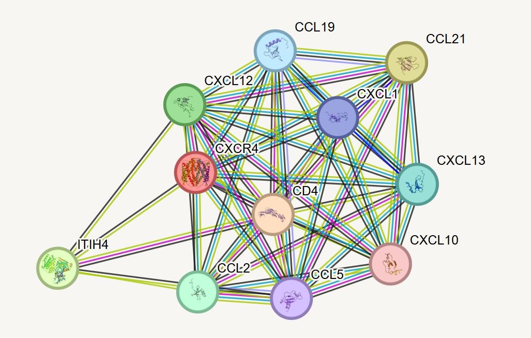
The STRING functional interaction network connected CXCR4 to a dense cluster of chemokines and receptors, including CXCL12, CCL5, CXCL10, CXCL13, and CD4. This confirms that CXCR4 functions within a coordinated multicellular immune trafficking network rather than in isolation.
Visual Bioinformatic Results
Limitations & Scientific Interpretation
All analyses were performed in silico using computational platforms and public secondary datasets. Association and correlation must be strictly distinguished from biological causation.
- Absence of Protein Validation: Analysis was restricted to mRNA transcript abundance; protein-level expression and receptor localization (e.g., cell surface versus inactive nuclear compartments) could not be determined without immunohistochemistry or Western blotting.
- Algorithmic Immune Deconvolution: TIMER2.0 estimates immune cell infiltration statistically from bulk transcriptomic profiles rather than physical cell sorting (flow cytometry); findings reflect cellular associations rather than demonstrated migration mechanisms.
- Automated Cutoff Thresholds: Survival curves utilized automated cutoff selection in Kaplan-Meier Plotter to optimize separation; validation across prospective clinical trials with fixed clinical thresholds is necessary.
- Public Dataset Heterogeneity: Findings rely on data processing standards within TCGA and GTEx, which are subject to technical batch variations, tumor stage distributions, and clinical treatment histories.
Key References
- Chatterjee, S., Behnam Azad, B., & Nimmagadda, S. (2014). The intricate role of CXCR4 in cancer. Advances in Cancer Research, 124, 31–82. DOI: 10.1016/B978-0-12-411638-2.00002-1
- Domanska, U. M. et al. (2013). A review on CXCR4/CXCL12 axis in oncology: No place to hide. European Journal of Cancer, 49(1), 219–230. DOI: 10.1016/j.ejca.2012.05.005
- Furusato, B., Mohamed, A., Uhlén, M., & Rhim, J. S. (2010). CXCR4 and cancer. Pathology International, 60(7), 497–505. DOI: 10.1111/j.1440-1827.2010.02548.x
- Liu, K. et al. (2015). Expression of CXCR4 and non-small cell lung cancer prognosis: A meta-analysis. Int J Clin Exp Med, 8(5), 7435–7445. PMC: PMC4503038
- Luker, G. D. et al. (2021). At the Bench: Pre-clinical evidence for multiple functions of CXCR4 in cancer. Journal of Leukocyte Biology, 109(5), 969–989. DOI: 10.1002/JLB.2BT1018-715RR
- Minamiya, Y. et al. (2010). Expression of the chemokine receptor CXCR4 correlates with a favorable prognosis in patients with adenocarcinoma of the lung. Lung Cancer, 68(3), 466–471. DOI: 10.1016/j.lungcan.2009.07.015
- Ottaiano, A. et al. (2021). Prognostic Significance of CXCR4 in Colorectal Cancer: An Updated Meta-Analysis. Cancers, 13(13), 3284. DOI: 10.3390/cancers13133284
- Spano, J.-P. et al. (2004). Chemokine receptor CXCR4 and early-stage non-small cell lung cancer. Annals of Oncology, 15(4), 613–617. DOI: 10.1093/annonc/mdh136
- Wagner, P. L. et al. (2009). CXCL12 and CXCR4 in adenocarcinoma of the lung: Association with metastasis and survival. J Thorac Cardiovasc Surg, 137(3), 615–621. DOI: 10.1016/j.jtcvs.2008.07.039
- Wald, O. et al. (2006). CD4+CXCR4highCD69+ T cells accumulate in lung adenocarcinoma. Journal of Immunology, 177(10), 6983–6990. DOI: 10.4049/jimmunol.177.10.6983
- Xu, C., Zhao, H., Chen, H., & Yao, Q. (2015). CXCR4 in breast cancer: Oncogenic role and therapeutic targeting. Drug Des Devel Ther, 9, 4953–4964. DOI: 10.2147/DDDT.S84932
- Zlotnik, A. (2008). New insights on the role of CXCR4 in cancer metastasis. The Journal of Pathology, 215(3), 211–213. DOI: 10.1002/path.2350